Article

Intravascular Ultrasound-guided Versus Angiography-guided Percutaneous Coronary Intervention: Evidence from Observational Studies and Randomized Controlled Trials

Register or Login to View PDF Permissions
Permissions× For commercial reprint enquiries please contact Springer Healthcare: ReprintsWarehouse@springernature.com.

For permissions and non-commercial reprint enquiries, please visit Copyright.com to start a request.

For author reprints, please email rob.barclay@radcliffe-group.com.
Information image
Average (ratings)
No ratings
Your rating

Abstract

Coronary angiography has been considered the gold standard for the diagnosis of coronary artery disease and guidance of percutaneous coronary intervention (PCI). However, 2D-projection angiography cannot completely reflect the 3D coronary lumen. Intravascular ultrasound (IVUS) can overcome a number of limitations of coronary angiography by providing more information about the dimensions of the vessel lumen, plaque characteristics, stent deployment, and the mechanisms of device failure. Growing data from observational studies and randomized controlled trials have confirmed the clinical benefit of IVUS guidance during PCI. This article summarizes the evidence regarding IVUS guidance to highlight its advantages and to support the use of IVUS during PCI.

Disclosure:The authors have no conflicts of interest to declare.

Received:

Accepted:

Published online:

Correspondence Details:Jun-Jie Zhang, MD, FSCAI, Department of Cardiology, Nanjing First Hospital, Nanjing Medical University; 68 Changle Rd, 210006 Nanjing, China. E: jameszll@163.com

Copyright Statement:

This work is open access under the CC-BY-NC 4.0 License which allows users to copy, redistribute and make derivative works for non-commercial purposes, provided the original work is cited correctly.

Coronary angiography is widely used to diagnose coronary artery disease and to guide percutaneous coronary intervention (PCI). However, 2D projection angiography cannot completely reflect the 3D coronary lumen, with several inherent limitations in evaluating plaque composition, vessel diameter, diffuse reference vessel disease, lesion severity, as well as the result of stent deployment. In the past three decades, intravascular ultrasound (IVUS) has been increasingly used in clinical practice to overcome a number of limitations of coronary angiography by providing more details of coronary anatomy and stent implantation.

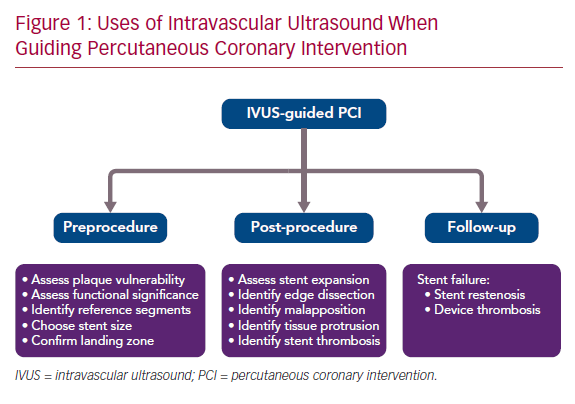
There is growing data from observational studies and randomized controlled trials (RCTs) to validate the value of IVUS guidance in PCI.1–19 IVUS guidance is not routinely performed in PCI, partly due to the increased procedural time, extra cost, and the potential neutral effect on cardiac death. This article summarizes the evidence for IVUS guidance in preprocedural, post-procedural, and follow-up assessment of PCI to highlight the advantage of using IVUS for patients undergoing stent implantation (Figure 1).

Preprocedural Assessment

Basic IVUS Measurement

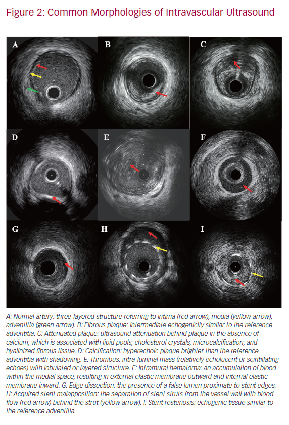
There are three layers in the ultrasound image of coronary arteries (Figure 2).20 The inner layer frequently includes atheroma, intima, and the internal elastic membrane. The middle layer is the media, which is less echogenic than the intima. The outer layer, bordering the external elastic membrane (EEM), consists of the adventitia and periadventitial tissues, which cannot be distinguished from each other in IVUS images. IVUS can be used to make the following basic measurements:

  • Minimum lumen diameter (MLD): the shortest diameter through the center point of the lumen.
  • Minimum lumen area (MLA): the smallest area through the center point of the lumen.
  • Lumen eccentricity: (maximum lumen diameter − minimum lumen diameter)/maximum lumen diameter.
  • Area stenosis: (reference lumen area − stenosis lumen area)/reference lumen area.
  • Plaque burden: (EEM area − lumen area)/EEM area.20

Assessment of Plaque Vulnerability

Vulnerable plaque, sometimes called thin-cap fibroatheromas (TCFA), often associated with large plaque burden, spotty calcifications, attenuated plaque, and shallow echolucent zones shown by gray-scale IVUS, is a common cause of MI and cardiac death.21–25 In virtual histology (VH)-IVUS, TCFA was defined as focal, necrotic core-rich plaque (≥10% of the cross-sectional area) in contact with the lumen with atheroma volume ≥40%.26,27 The international, multicenter trial An Imaging Study in Patients with Unstable Atherosclerotic Lesions (PROSPECT) enrolled 697 patients with acute coronary syndrome undergoing coronary angiography, gray-scale and radiofrequency IVUS. It showed that three lesion-level independent predictors of major adverse cardiovascular events (MACE) at a median follow-up of 3.4 years were MLA ≤4 mm2 (HR 3.21; 95% CI [1.61–6.42]), plaque burden ≥70% (HR 5.03; 95% CI [2.51–10.11]), and the presence of TCFA (HR 3.35; 95% CI [1.77–6.36]).28 In this study, TCFA was defined as the presence of >10% confluent necrotic core with more than 30% of the necrotic core abutting the lumen in ≥3 consecutive frames by VH-IVUS. Moreover, the VH-IVUS in Vulnerable Atherosclerosis (VIVA) study and the European Collaborative Project on Inflammation and Vascular Wall Remodeling in Atherosclerosis – Intravascular Ultrasound (ATHEROREMO-IVUS) study confirmed the finding that TCFA identified by VH-IVUS was associated with MACE.27,29 However, the PROSPECT trial also showed that the combination of these three high-risk plaque features only had an 18.2% predictive value for MACE, which might be due to the low resolution of IVUS, which makes it difficult to detect plaque composition.28

Uses of Intravascular Ultrasound When Guiding Percutaneous Coronary Intervention

Article image

Common Morphologies of Intravascular Ultrasound

Article image

Assessment of Functional Significance

There is always high inter-observer variability for angiographic estimation of the degree of coronary stenosis.30,31 Intermediate coronary lesions, defined by 40–70% stenosis by angiography assessment, cannot be accurately evaluated for their hemodynamic significance for MI even by experienced interventional cardiologists. Fractional flow reserve (FFR) has been regarded as the gold standard method of invasive MI assessment.32–34 Anatomic data for MLA has a relatively good correlation with FFR, which could be a liberal diagnostic application, though potential errors exist due to the variations of BMI and lesion complexity.

A comprehensive meta-analysis demonstrated that an IVUS-derived MLA of 2.8 mm2 for non-left main coronary lesions with an angiographic diameter >3 mm, and 2.4 mm2 for lesions with a diameter <3 mm, were cut-off values to detect functionally significant coronary stenosis.35 In isolated intermediate left main coronary lesions involving the ostium or shaft, an IVUS-derived MLA of 5.9 mm2 had the highest sensitivity (93%) and specificity (95%) for determining FFR less than 0.75 in Western populations, while an MLA <4.5 mm2 had good correlation with an FFR under 0.80 (77% sensitivity and 82% specificity) in Asian populations.36,37

The multicenter, prospective Spanish Working Group on Interventional Cardiology (LITRO) study also showed that an IVUS-derived MLA of more than 6 mm2 was a safe value to use to defer coronary revascularization of the left main lesions.38 Therefore, it seems reasonable to perform revascularization for left main coronary lesions when there is an MLA of <4.5 mm2, to defer revascularization if there is an MLA of >6 mm2 and to consider further invasive or non-invasive functional evaluation if there is an MLA of 4.5–6 mm2.

IVUS-guided Preprocedural Preparation

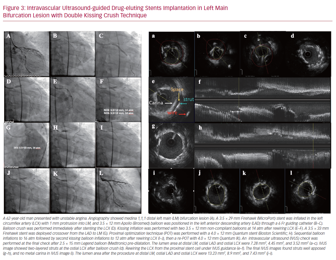
A preprocedural IVUS check is important to assess calcium severity, to select stent size, to identify a reference segment and confirm the landing zone (Figure 3). Angiography is moderately sensitive for the detection of extensive calcific lesions, but it is less sensitive for mild calcium. One study has shown that IVUS could detect calcium in 841 of 1,155 (73%) stable patients, whereas angiography detected calcium in only 440 (38%) of them.39 The presence of severe calcium may require predilatation with a higher inflation pressure, larger balloon, cutting balloon angioplasty, or rotablator atherectomy.

Several potential IVUS-based stent diameter methods exist, including stent diameter according to EEM diameter at the site of MLA, the smallest reference EEM diameter, the largest reference lumen, mean reference lumen, or the smallest reference lumen. Currently, the use of mean diameter of distal lumen with post-dilatation of the proximal and middle part of the stent is recommended.40 Stent length is determined by the distance from the distal to proximal reference site. Proximal and distal reference sites are set at cross-sections adjacent to the target lesion that have the largest lumen and a plaque burden of <50%.41 An appropriate landing zone is commonly considered as having residual plaque burden <50% without lipid-rich plaque at the stent edge, which is associated with subsequent stent restenosis.40–42 Overall, compared with angiography guidance, IVUS guidance could lead to more stents, larger stent diameter, longer stent length, and a greater post-procedural minimum stent area (MSA).12,19,43-45

Intravascular Ultrasound-guided Drug-eluting Stents Implantation

Article image

Post-procedural Assessment

Post-procedural IVUS assessment can detect stent underexpansion, acute stent malapposition, stent deformation, tissue protrusion through the stent struts, stent edge dissection, and residual disease at stent edge (Figures 2 and 3). Several studies have demonstrated that stent underexpansion was associated with early stent thrombosis and restenosis after implantation of a drug-eluting stent (DES).46–49 Post-procedural MSA has been considered the most important parameter to predict these adverse events.46–51 The current view is that the optimal MSA measured by IVUS is >5.5 mm2 for non-left main lesions, >7 mm2 for distal left main lesions, and >8 mm2 for proximal left main lesions.40,47,48,52,53 Acute stent malapposition after DES implantation without underexpansion does not translate into early or long-term adverse events regardless of the length and thickness of malapposition.54–56

An IVUS subgroup analysis from the Assessment of Dual AntiPlatelet Therapy With Drug Eluting Stents (ADAPT-DES) trial showed that 34.3% of lesions presented with tissue protrusion detected by IVUS after DES implantation. This was not associated with worse 2-year clinical outcomes, in part due to the larger lumen area of lesions treated with larger stent or post-dilatation balloon in the tissue protrusion group.57

However, a substudy of the Bivalirudin in Patients Undergoing Primary Angioplasty for Acute Myocardial Infarction (HORIZONS-AMI) trial demonstrated that significant tissue protrusion (plaque/thrombus), defined as residual lumen area <4mm2 by IVUS detection, was associated with early stent thrombosis.46 Moreover, post-procedural large edge dissections – characterized by IVUS as deep depth (at least disrupting the media layer) great lateral extension (>60°C), and long length (>2 mm) – could result in early stent thrombosis.46,58

Key Randomized Trials of Intravascular Ultrasound

Article image

The optimal criteria of IVUS-guided bare-metal stent deployment in the Multicenter Ultrasound Stenting in Coronaries Study (MUSIC) included:

  • complete apposition of stent;
  • adequate stent expansion: MSA ≥90% of average reference lumen area or ≥100% of the smaller reference segment area if the MSA <9 mm2, or MSA ≥80% of average reference lumen area or ≥90% of the smaller reference segment area if the MSA >9 mm2; and
  • symmetrical stent expansion: MLD/maximum lumen diameter ≥0.7.

The MUSIC study found that 81% of 155 patients undergoing bare metal Palmaz-Schatz stents met IVUS optimal criteria, and the overall risk of target lesion revascularization was 4.5% at 6 months.59

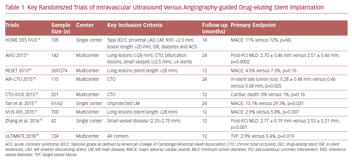
In the DES era, evidence derived from observational studies, RCTs (Table 1) and meta-analyses all demonstrated that IVUS-guided DES implantation was associated with a lower risk of MACE and target vessel revascularization (TVR) in complex lesions, such as unprotected left main disease, bifurcation lesions, chronic total occlusion, and long lesions.1–5,8,10–18,43,44,60–65 More importantly, several optimal IVUS-guided criteria have been proposed through RCTs rather than observational studies. The Angiography Versus (vs) IVUS Optimisation (AVIO) RCT, proposed a new optimal IVUS-guided criterion: MSA >70% of the post-dilatation balloon cross-sectional area (CSA), and the non-compliant post-dilatation balloon size to be determined by the average of the media-to-media diameters of distal in-stent segment, proximal in-stent segment, and maximal in-stent narrowing.11

Another randomized trial – the Impact of Intravascular Ultrasound Guidance on Outcomes of Xience Prime Stents in Long Lesions (IVUS-XPL) study – presented an IVUS-guided optimal criterion for long lesions as the MLA greater than the lumen CSA at distal reference segments.12,65 Most RCTs have enrolled people with complex coronary lesions, and only the Intravascular Ultrasound Guided Drug Eluting Stents Implantation in ‘All-Comers’ Coronary Lesions (ULTIMATE) trial recruited all-comer patients and showed that IVUS guidance was associated with significant lower risk of target vessel failure (TVF) compared with angiography guidance in all-comers undergoing second-generation DES implantation.19 In this trial, the novel criteria of IVUS-guided optimal DES deployment were:

  • the MLA in the stented segment >5.0 mm2, or 90% of the MLA at the distal reference segments;
  • plaque burden of 5 mm proximal or distal to the stent edge <50%; and
  • no edge dissection involving media with a length >3 mm.

A total of 53% of the 1,448 participants met these three criteria and they were associated with a lower rate of TVF at 12 months compared with those with a suboptimal PCI procedure. An updated meta-analysis that included the ULTIMATE trial demonstrated that IVUS guidance could reduce the risk of cardiac death.66 A 5-year clinical follow-up of the IVUS-XPL trial has shown the long-term benefit of IVUS guidance in optimizing DES implantation in long lesions.65 However, further RCTs are warranted to explore the difference in clinical relevance when using different optimal IVUS guidance criteria.67

Follow-up Assessment

IVUS at follow-up is used to detect chronic stent expansion, stent fracture, neointimal hyperplasia, stent malapposition, and positive remodeling of vessel wall. Current guidelines and expert consensus recommend intracoronary imaging should be used to identify the mechanisms of stent failure (restenosis and thrombosis) at follow-up.32,40,68

The common causes of stent restenosis, apart from intimal hyperplasia, are chronic underexpansion, stent fracture, and neoatherosclerosis.47,69 Chronic underexpansion and stent fracture could be assessed easily by IVUS, but the detection of neoatherosclerosis may need a higher resolution intracoronary modality. A prospective, multicenter study showed that stent fracture could be found in 803 (12.3%) patients, 3,630 (22.0%) stents, and 1,852 (17.2%) diseased vessels, which was associated with higher risk of stent restenosis and definite stent thrombosis.70 In this study, a novel classification of stent fracture was proposed, identifying five types:

  • type IA: single strut fracture;
  • type IB: gap between 2 struts >2 times a 2.5mm cell;
  • type II: incomplete transverse, V gap;
  • type III: complete transverse, no displacement; and
  • type IV: complete transverse with displacement.

Of 3,630 fractured stents in this study 2,963 were detected by angiography and the remaining 640 had to be identified by IVUS. Stent malapposition at follow-up should be divided into two types: persistent malapposition since stent implantation and late acquired malapposition, which may be caused by plaque/thrombus resolution and positive remodeling.54

Conclusion

IVUS can provide important information about vessel lumen, dimensions, plaque characteristics, and stent deployment, as well as the mechanisms of device failure. Clinical studies have demonstrated that IVUS-guided PCI could improve the clinical outcomes in patients with DES implantation, especially for complex coronary lesions and high-risk patients. But IVUS guidance is not routinely performed in the real-world daily practice of PCI, partly due to the increased procedural time and extra cost. The next step should be to reduce the cost of IVUS, educate interventional cardiologists, and promote the use of IVUS as much as possible during PCI.

References

  1. Chen L, Xu T, Xue XJ, et al. Intravascular ultrasound-guided drug-eluting stent implantation is associated with improved clinical outcomes in patients with unstable angina and complex coronary artery true bifurcation lesions. Int J Cardiovasc Imaging 2018; 34:1685–96.
    Crossref | PubMed
  2. Chen SL, Ye F, Zhang JJ, et al. Intravascular ultrasound-guided systematic two-stent techniques for coronary bifurcation lesions and reduced late stent thrombosis. Catheter Cardiovasc Interv 2013;81:456–63.
    Crossref | PubMed
  3. Gao XF, Kan J, Zhang YJ, et al. Comparison of one-year clinical outcomes between intravascular ultrasound-guided versus angiography-guided implantation of drug-eluting stents for left main lesions: a single-center analysis of a 1,016-patient cohort. Patient Prefer Adherence 2014;8:1299–1309.
    Crossref | PubMed
  4. Andell P, Karlsson S, Mohammad MA, et al. Intravascular ultrasound guidance is associated with better outcome in patients undergoing unprotected left main coronary artery stenting compared with angiography guidance alone. Circ Cardiovasc Interv 2017;10:pii:e004813.
    Crossref | PubMed
  5. Choi KH, Song YB, Lee JM, et al. Impact of intravascular ultrasound-guided percutaneous coronary intervention on long-term clinical outcomes in patients undergoing complex procedures. JACC Cardiovasc Interv 2019;12:607–20.
    Crossref | PubMed
  6. Frohlich GM, Redwood S, Rakhit R, et al. Long-term survival in patients undergoing percutaneous interventions with or without intracoronary pressure wire guidance or intracoronary ultrasonographic imaging: a large cohort study. JAMA Intern Med 2014;174:1360–6.
    Crossref | PubMed
  7. Park KW, Kang SH, Yang HM, et al. Impact of intravascular ultrasound guidance in routine percutaneous coronary intervention for conventional lesions: data from the EXCELLENT trial. Int J Cardiol 2013;167:721–6.
    Crossref | PubMed
  8. Park SJ, Kim YH, Park DW, et al. Impact of intravascular ultrasound guidance on long-term mortality in stenting for unprotected left main coronary artery stenosis. Circ Cardiovasc Interv 2009;2:167–77.
    Crossref | PubMed
  9. Witzenbichler B, Maehara A, Weisz G, et al. Relationship between intravascular ultrasound guidance and clinical outcomes after drug-eluting stents: the assessment of dual antiplatelet therapy with drug-eluting stents (ADAPT-DES) study. Circulation 2014;129:463–70.
    Crossref | PubMed
  10. Shlofmitz E, Torguson R, Zhang C, et al. Impact of Intravascular ultrasound on Outcomes following PErcutaneous coronary interventioN in Complex Lesions (iOPEN Complex). Am Heart J 2019;221:74–83.
    Crossref | PubMed
  11. Chieffo A, Latib A, Caussin C, et al. A prospective, randomized trial of intravascular-ultrasound guided compared to angiography guided stent implantation in complex coronary lesions: the AVIO trial. Am Heart J 2013;165:65–72.
    Crossref | PubMed
  12. Hong SJ, Kim BK, Shin DH, et al. Effect of intravascular ultrasound-guided vs angiography-guided everolimus-eluting stent implantation: the IVUS-XPL randomized clinical trial. JAMA 2015;314:2155–63.
    Crossref | PubMed
  13. Jakabcin J, Spacek R, Bystron M, et al. Long-term health outcome and mortality evaluation after invasive coronary treatment using drug eluting stents with or without the IVUS guidance. Randomized control trial. HOME DES IVUS. Catheter Cardiovasc Interv 2010; 75:578–83.
    Crossref | PubMed
  14. Zhang JQ, Pang WP, Guo Q, et al. Application of intravascular ultrasound in stent implantation for small coronary arteries. Journal of Clinical & Invasive Cardiology 2016;3:2–8.
  15. Kim BK, Shin DH, Hong MK, et al. Clinical impact of intravascular ultrasound-guided chronic total occlusion intervention with zotarolimus-eluting versus biolimus-eluting stent implantation: randomized study. Circ Cardiovasc Interv 2015;8:e002592.
    Crossref | PubMed
  16. Kim JS, Kang TS, Mintz GS, et al. Randomized comparison of clinical outcomes between intravascular ultrasound and angiography-guided drug-eluting stent implantation for long coronary artery stenoses. JACC Cardiovasc Interv 2013;6:369–76.
    Crossref | PubMed
  17. Tan Q, Wang Q, Liu D, et al. Intravascular ultrasound-guided unprotected left main coronary artery stenting in the elderly. Saudi Med J 2015;36:549–53.
    Crossref | PubMed
  18. Tian NL, Gami SK, Ye F, et al. Angiographic and clinical comparisons of intravascular ultrasound- versus angiography-guided drug-eluting stent implantation for patients with chronic total occlusion lesions: two-year results from a randomised AIR-CTO study. EuroIntervention 2015;10:1409–17.
    Crossref | PubMed
  19. Zhang J, Gao X, Kan J, et al. Intravascular ultrasound versus angiography-guided drug-eluting stent implantation: the ULTIMATE Trial. J Am Coll Cardiol 2018;72:3126–37.
    Crossref | PubMed
  20. Mintz GS, Nissen SE, Anderson WD, et al. American College of Cardiology clinical expert consensus document on standards for acquisition, measurement and reporting of intravascular ultrasound studies (IVUS). A report of the American College of Cardiology Task Force on Clinical Expert Consensus Documents. J Am Coll Cardiol 2001;37:1478–92.
    Crossref | PubMed
  21. Virmani R, Burke AP, Farb A, Kolodgie FD. Pathology of the vulnerable plaque. J Am Coll Cardiol 2006;47:C13–8.
    Crossref | PubMed
  22. Naghavi M, Libby P, Falk E, et al. From vulnerable plaque to vulnerable patient: a call for new definitions and risk assessment strategies: Part I. Circulation 2003;108:1664–72.
    Crossref | PubMed
  23. Naghavi M, Libby P, Falk E, et al. From vulnerable plaque to vulnerable patient: a call for new definitions and risk assessment strategies: part II. Circulation 2003;108:1772–8.
    Crossref | PubMed
  24. Garcia-Garcia HM, Mintz GS, Lerman A, et al. Tissue characterisation using intravascular radiofrequency data analysis: recommendations for acquisition, analysis, interpretation and reporting. EuroIntervention 2009;5:177–89.
    Crossref | PubMed
  25. Burke AP, Farb A, Malcom GT, et al. Coronary risk factors and plaque morphology in men with coronary disease who died suddenly. N Engl J Med 1997;336:1276–82.
    Crossref | PubMed
  26. Rodriguez-Granillo GA, Garcia-Garcia HM, Mc Fadden EP, et al. In vivo intravascular ultrasound-derived thin-cap fibroatheroma detection using ultrasound radiofrequency data analysis. J Am Coll Cardiol 2005;46:2038–42.
    Crossref | PubMed
  27. Calvert PA, Obaid DR, O’Sullivan M, et al. Association between IVUS findings and adverse outcomes in patients with coronary artery disease: the VIVA (VH-IVUS in Vulnerable Atherosclerosis) Study. JACC Cardiovasc Imaging 2011;4:894–901.
    Crossref | PubMed
  28. Stone GW, Maehara A, Lansky AJ, et al. A prospective natural-history study of coronary atherosclerosis. N Engl J Med 2011;364:226–35.
    Crossref | PubMed
  29. Cheng JM, Garcia-Garcia HM, de Boer SP, et al. In vivo detection of high-risk coronary plaques by radiofrequency intravascular ultrasound and cardiovascular outcome: results of the ATHEROREMO-IVUS study. Eur Heart J 2014;35:639–47.
    Crossref | PubMed
  30. Fischer JJ, Samady H, McPherson JA, et al. Comparison between visual assessment and quantitative angiography versus fractional flow reserve for native coronary narrowings of moderate severity. Am J Cardiol 2002;90:210–5.
    Crossref | PubMed
  31. Jensen LO, Thayssen P, Mintz GS, et al. Comparison of intravascular ultrasound and angiographic assessment of coronary reference segment size in patients with type 2 diabetes mellitus. Am J Cardiol 2008;101:590–5.
    Crossref | PubMed
  32. Neumann FJ, Sousa-Uva M, Ahlsson A, et al. 2018 ESC/EACTS Guidelines on myocardial revascularization. Eur Heart J 2019;40:87–165.
    Crossref | PubMed
  33. De Bruyne B, Pijls NH, Kalesan B, et al. Fractional flow reserve-guided PCI versus medical therapy in stable coronary disease. N Engl J Med 2012;367:991–1001.
    Crossref | PubMed
  34. Tonino PA, De Bruyne B, Pijls NH, et al. Fractional flow reserve versus angiography for guiding percutaneous coronary intervention. N Engl J Med 2009;360:213–24.
    Crossref | PubMed
  35. D’Ascenzo F, Barbero U, Cerrato E, et al. Accuracy of intravascular ultrasound and optical coherence tomography in identifying functionally significant coronary stenosis according to vessel diameter: a meta-analysis of 2,581 patients and 2,807 lesions. Am Heart J 2015;169:663–73.
    Crossref | PubMed
  36. Jasti V, Ivan E, Yalamanchili V, et al. Correlations between fractional flow reserve and intravascular ultrasound in patients with an ambiguous left main coronary artery stenosis. Circulation 2004;110:2831–6.
    Crossref | PubMed
  37. Park SJ, Ahn JM, Kang SJ, et al. Intravascular ultrasound-derived minimal lumen area criteria for functionally significant left main coronary artery stenosis. JACC Cardiovasc Interv 2014;7:868–74.
    Crossref | PubMed
  38. de la Torre Hernandez JM, Hernandez Hernandez F, Alfonso F, et al. Prospective application of pre-defined intravascular ultrasound criteria for assessment of intermediate left main coronary artery lesions: results from the multicenter LITRO study. J Am Coll Cardiol 2011;58:351–8.
    Crossref | PubMed
  39. Mintz GS, Popma JJ, Pichard AD, et al. Patterns of calcification in coronary artery disease. A statistical analysis of intravascular ultrasound and coronary angiography in 1155 lesions. Circulation 1995;91:1959–65.
    Crossref | PubMed
  40. Raber L, Mintz GS, Koskinas KC, et al. Clinical use of intracoronary imaging. Part 1: guidance and optimization of coronary interventions. An expert consensus document of the European Association of Percutaneous Cardiovascular Interventions. Eur Heart J 2018;39: 3281–300.
    Crossref | PubMed
  41. Liu J, Maehara A, Mintz GS, et al. An integrated TAXUS IV, V, and VI intravascular ultrasound analysis of the predictors of edge restenosis after bare metal or paclitaxel-eluting stents. Am J Cardiol 2009;103:501–6.
    Crossref | PubMed
  42. Kang SJ, Cho YR, Park GM, et al. Intravascular ultrasound predictors for edge restenosis after newer generation drug-eluting stent implantation. Am J Cardiol 2013;111:1408–14.
    Crossref | PubMed
  43. Elgendy IY, Mahmoud AN, Elgendy AY, Bavry AA. Outcomes with intravascular ultrasound-guided stent implantation: a meta-analysis of randomized trials in the era of drug-eluting stents. Circ Cardiovasc Interv 2016;9:e003700.
    Crossref | PubMed
  44. Bavishi C, Sardar P, Chatterjee S, et al. Intravascular ultrasound-guided vs angiography-guided drug-eluting stent implantation in complex coronary lesions: Meta-analysis of randomized trials. Am Heart J 2017;185:26–34.
    Crossref | PubMed
  45. Ahn JM, Kang SJ, Yoon SH, et al. Meta-analysis of outcomes after intravascular ultrasound-guided versus angiography-guided drug-eluting stent implantation in 26,503 patients enrolled in three randomized trials and 14 observational studies. Am J Cardiol 2014;113:1338–47.
    Crossref | PubMed
  46. Choi SY, Witzenbichler B, Maehara A, et al. Intravascular ultrasound findings of early stent thrombosis after primary percutaneous intervention in acute myocardial infarction: a Harmonizing Outcomes with Revascularization and Stents in Acute Myocardial Infarction (HORIZONS-AMI) substudy. Circ Cardiovasc Interv 2011;4:239–47.
    Crossref | PubMed
  47. Kang SJ, Ahn JM, Song H, et al. Comprehensive intravascular ultrasound assessment of stent area and its impact on restenosis and adverse cardiac events in 403 patients with unprotected left main disease. Circ Cardiovasc Interv 2011;4:562–9.
    Crossref | PubMed
  48. Song HG, Kang SJ, Ahn JM, et al. Intravascular ultrasound assessment of optimal stent area to prevent in-stent restenosis after zotarolimus-, everolimus-, and sirolimus-eluting stent implantation. Catheter Cardiovasc Interv 2014;83:873–8.
    Crossref | PubMed
  49. Sonoda S, Morino Y, Ako J, et al. Impact of final stent dimensions on long-term results following sirolimus-eluting stent implantation: serial intravascular ultrasound analysis from the sirius trial. J Am Coll Cardiol 2004;43:1959–63.
    Crossref | PubMed
  50. Liu X, Doi H, Maehara A, et al. A volumetric intravascular ultrasound comparison of early drug-eluting stent thrombosis versus restenosis. JACC Cardiovasc Interv 2009;2:428–34.
    Crossref | PubMed
  51. Moussa I, Moses J, Di Mario C, et al. Does the specific intravascular ultrasound criterion used to optimize stent expansion have an impact on the probability of stent restenosis? Am J Cardiol 1999;83:1012–7.
    Crossref | PubMed
  52. Doi H, Maehara A, Mintz GS, et al. Impact of post-intervention minimal stent area on 9-month follow-up patency of paclitaxel-eluting stents: an integrated intravascular ultrasound analysis from the TAXUS IV, V, and VI and TAXUS ATLAS Workhorse, Long Lesion, and Direct Stent Trials. JACC Cardiovasc Interv 2009;2:1269–75.
    Crossref | PubMed
  53. Hong MK, Mintz GS, Lee CW, et al. Intravascular ultrasound predictors of angiographic restenosis after sirolimus-eluting stent implantation. Eur Heart J 2006;27:1305–10.
    Crossref | PubMed
  54. Guo N, Maehara A, Mintz GS, et al. Incidence, mechanisms, predictors, and clinical impact of acute and late stent malapposition after primary intervention in patients with acute myocardial infarction: an intravascular ultrasound substudy of the Harmonizing Outcomes with Revascularization and Stents in Acute Myocardial Infarction (HORIZONS-AMI) trial. Circulation 2010;122:1077–84.
    Crossref | PubMed
  55. Romagnoli E, Gatto L, La Manna A, et al. Role of residual acute stent malapposition in percutaneous coronary interventions. Catheter Cardiovasc Interv 2017;90:566–75.
    Crossref | PubMed
  56. Wang B, Mintz GS, Witzenbichler B, et al. Predictors and long-term clinical impact of acute stent malapposition: an assessment of dual antiplatelet therapy with drug-eluting stents (ADAPT-DES) intravascular ultrasound substudy. J Am Heart Assoc 2016;5:pii:e004438.
    Crossref | PubMed
  57. Qiu F, Mintz GS, Witzenbichler B, et al. Prevalence and clinical impact of tissue protrusion after stent implantation: an ADAPT-DES intravascular ultrasound substudy. JACC Cardiovasc Interv 2016;9:1499–1507.
    Crossref | PubMed
  58. Cheneau E, Leborgne L, Mintz GS, et al. Predictors of subacute stent thrombosis: results of a systematic intravascular ultrasound study. Circulation 2003;108:43–7.
    Crossref | PubMed
  59. de Jaegere P, Mudra H, Figulla H, et al. Intravascular ultrasound-guided optimized stent deployment. Immediate and 6 months clinical and angiographic results from the Multicenter Ultrasound Stenting in Coronaries Study (MUSIC Study). Eur Heart J 1998;19: 1214–23.
    Crossref | PubMed
  60. Zhang J, Gao X, Ge Z, et al. Impact of intravascular ultrasound-guided drug-eluting stent implantation on patients with chronic kidney disease: results from ULTIMATE trial. Catheter Cardiovasc Interv 2019;93:1184–93.
    Crossref | PubMed
  61. Buccheri S, Franchina G, Romano S, et al. Clinical outcomes following intravascular imaging-guided versus coronary angiography-guided percutaneous coronary intervention with stent implantation: a systematic review and bayesian network meta-analysis of 31 studies and 17,882 patients. JACC Cardiovasc Interv 2017;10:2488–98.
    Crossref | PubMed
  62. Shin DH, Hong SJ, Mintz GS, et al. Effects of intravascular ultrasound-guided versus angiography-guided new-generation drug-eluting stent implantation: meta-analysis with individual patient-level data from 2,345 randomized patients. JACC Cardiovasc Interv 2016;9:2232–9.
    Crossref | PubMed
  63. Steinvil A, Zhang YJ, Lee SY, et al. Intravascular ultrasound-guided drug-eluting stent implantation: an updated meta-analysis of randomized control trials and observational studies. Int J Cardiol 2016;216:133–9.
    Crossref | PubMed
  64. Fan ZG, Gao XF, Li XB, et al. The outcomes of intravascular ultrasound-guided drug-eluting stent implantation among patients with complex coronary lesions: a comprehensive meta-analysis of 15 clinical trials and 8,084 patients. Anatol J Cardiol 2017;17:258–68.
    Crossref | PubMed
  65. Hong SJ, Mintz GS, Ahn CM, et al. Effect of intravascular ultrasound-guided drug-eluting stent implantation: 5-year follow-up of the IVUS-XPL randomized trial. JACC Cardiovasc Interv 2020;13:62–71.
    Crossref | PubMed
  66. Gao XF, Wang ZM, Wang F, et al. Intravascular ultrasound guidance reduces cardiac death and coronary revascularization in patients undergoing drug-eluting stent implantation: results from a meta-analysis of 9 randomized trials and 4,724 patients. Int J Cardiovasc Imaging 2019;35:239–47.
    Crossref | PubMed
  67. Zhang JJ, Chen SL. IVUS guidance for coronary revascularization: when to start, when to stop? JACC Cardiovasc Interv 2020;13:72–3.
    Crossref | PubMed
  68. Johnson TW, Raber L, di Mario C, et al. Clinical use of intracoronary imaging. Part 2: acute coronary syndromes, ambiguous coronary angiography findings, and guiding interventional decision-making: an expert consensus document of the European Association of Percutaneous Cardiovascular Interventions. Eur Heart J 2019;40:2566–84.
    Crossref | PubMed
  69. Goto K, Zhao Z, Matsumura M, et al. Mechanisms and patterns of intravascular ultrasound in-stent restenosis among bare metal stents and first- and second-generation drug-eluting stents. Am J Cardiol 2015;116:1351–7.
    Crossref | PubMed
  70. Kan J, Ge Z, Zhang JJ, et al. Incidence and clinical outcomes of stent fractures on the basis of 6,555 patients and 16,482 drug-eluting stents from 4 centers. JACC Cardiovasc Interv 2016;9:1115–23.
    Crossref | PubMed